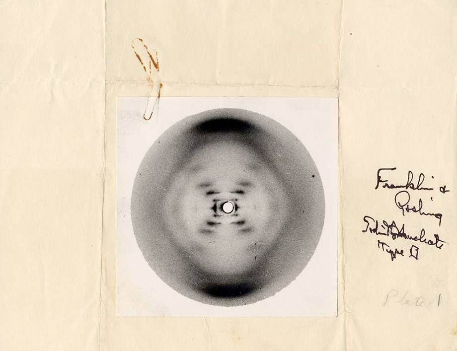
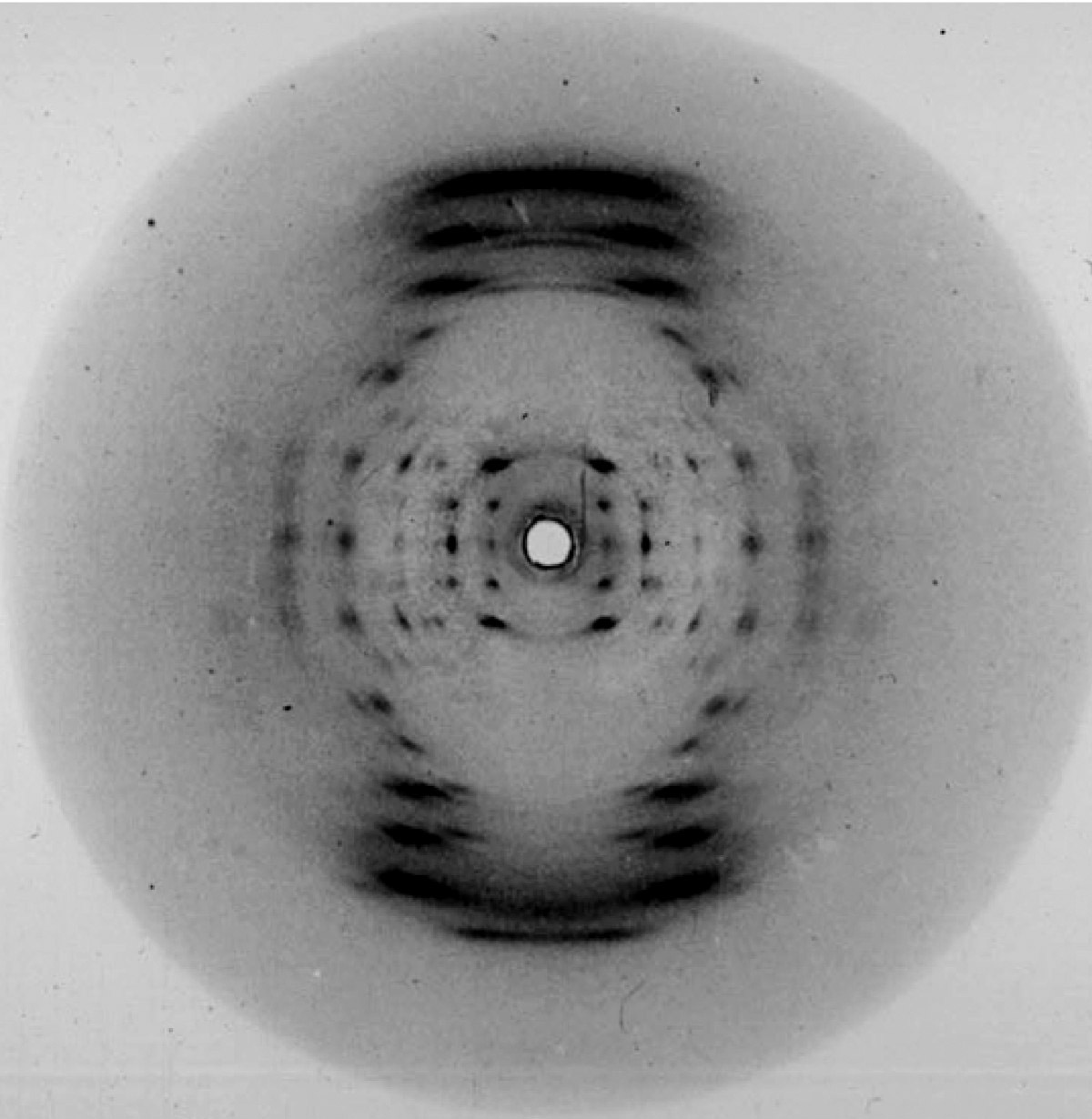
Linus Pauling's copy of Photo 51, 2 May 1953. Oregon State University Library
Photo 51
Rosalind Franklin and the Structure of DNA
Sometime in May 1952 Raymond Gosling, a doctoral candidate under Rosalind Franklin at the Wheatstone Physics Labratory, King’s College, took some 35 strands of sodium thymonucleate obtained from a calf thymus, stretched them taut across a paperclip mounted to a cork with rubber cement and sealed the makeshift assembly in a camera fitted to an X-ray tube. Gosling then bubbled water and hydrogen through the camera and exposed the film for nearly 90 hours. Later he recalled “looking at the developer, and up through the tank swam this beautiful spotted photograph.”
Franklin and Gosling’s “spotted photograph” – now known as simply Photo 51 – was to anyone who could interpret it the first clear evidence of the structure of DNA. It would become the most iconic photograph in the history of molecular biology.
In 1869 the Swiss physician Johannes Miescher first isolated and identified a molecule which he called “nuclein” in the pus of left over surgical bandages. Later researchers including the Lithuanian-American biochemist Phoebus Levene and the Austrian-American biochemist Erwin Chargroff determined the basic chemical composition of the molecule, which was now known as nucleic acid. In 1944 Oswald Avery at Rockefeller University conclusively determined that this molecule, now known as desoxyribonucleic acid (or deoxyribonucleic acid, or just DNA), was responsable for the transmission of genetic material.1
By this time researchers had figured out the composition and role of DNA, but its physical structure – and therefore how it worked – remained elusive, so scientists began to apply the relatively new method of X-ray diffraction to the problem. The first X-ray photos were from the lab of William Astbury at the University of Leeds:
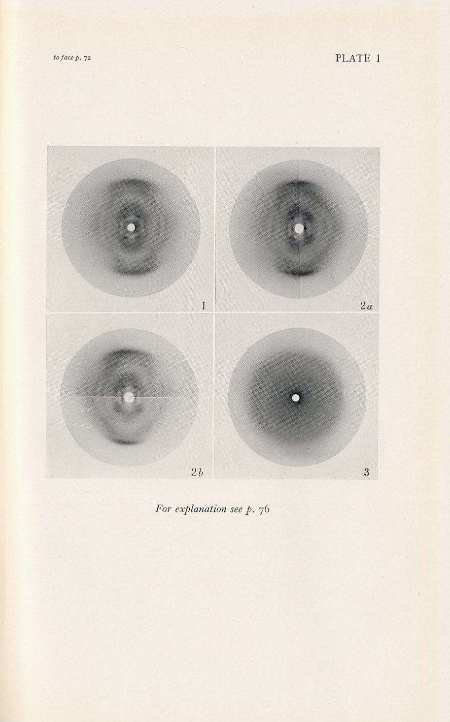
Sodium thymonucleate and clupein thymonucleate. From ref. 2
The physicist Maurice Wilkins at King’s college, who had spent the war on the Manhattan Project, produced this image using a Unicam camera and Raymax tube in 1950:
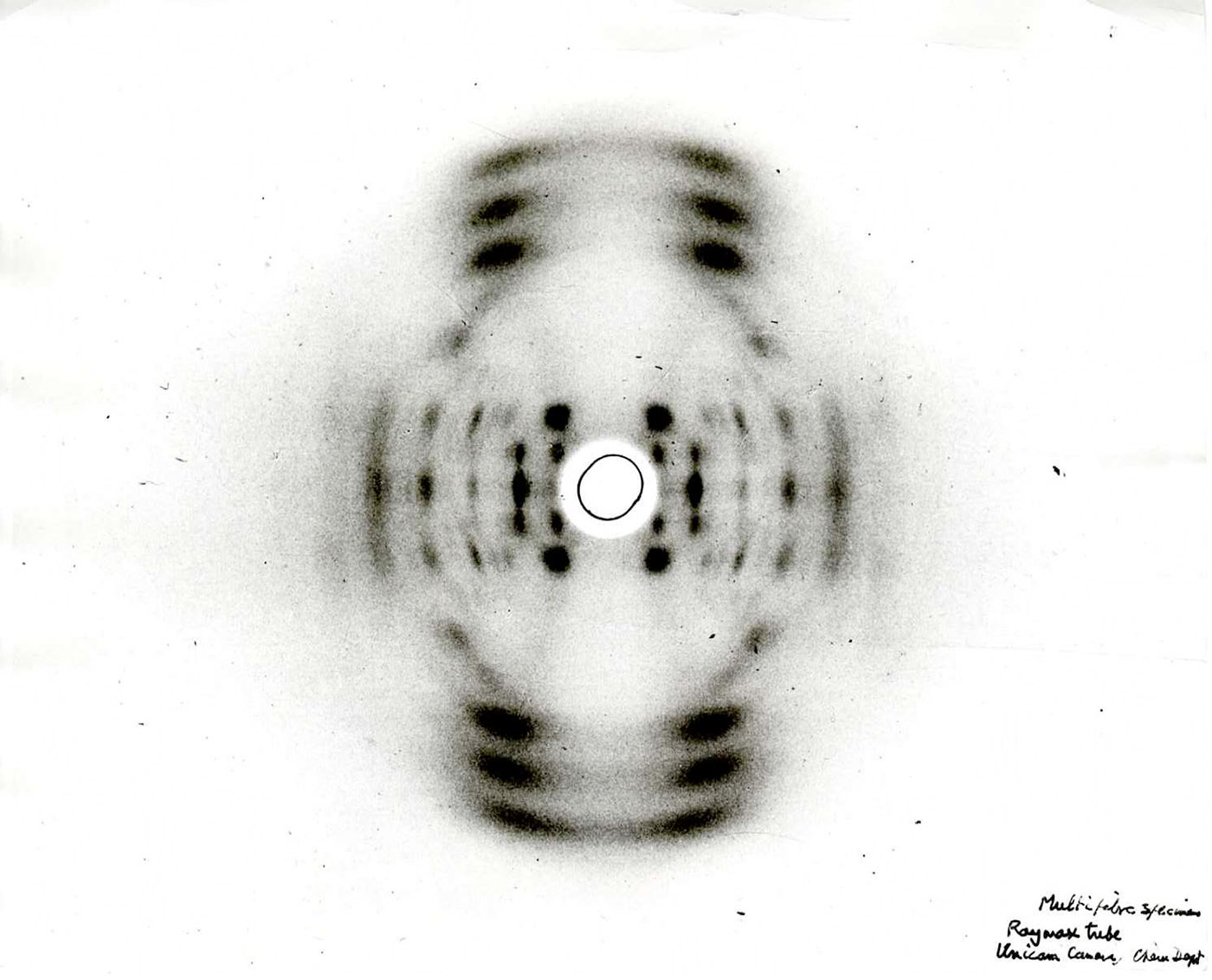
Wilkins and Gosling. Multifiber specimen, 1950. King's College Archive
Aside from the rather obvious fact that DNA was somehow symmetrical, these early photos did little to explain the structure. This is where Rosalind Franklin comes in.
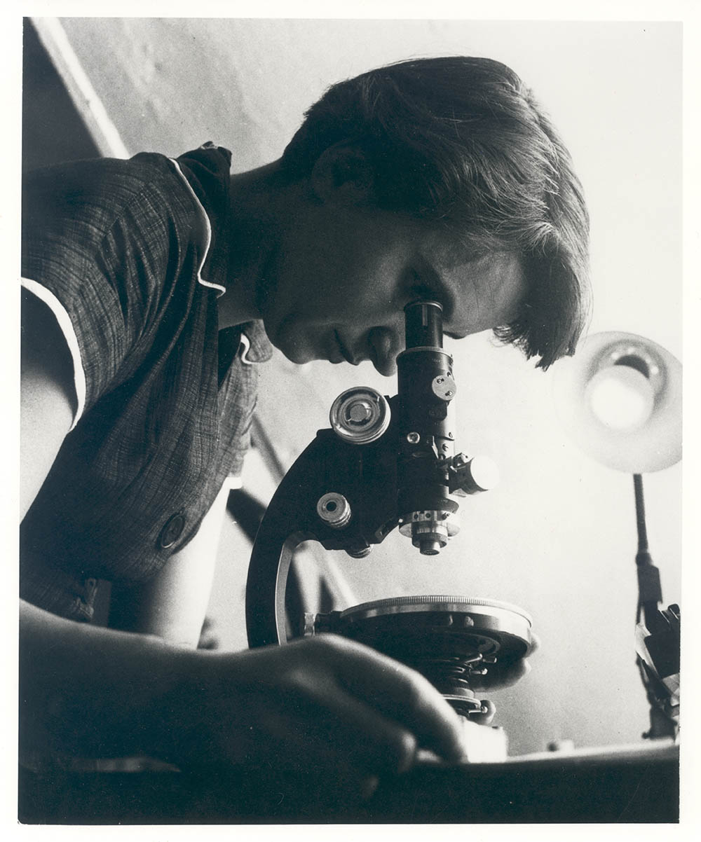
Rosalind at Birkbeck, 1955. National Library of Medicine
Franklin spent her childhood enjoying memory games and doing arithmetic for pleasure. As her aunt said “she was alarmingly clever.” It was clear that she was destined for a career in science and against her parents wishes, received an M.S. degree in chemistry from Newham College, Cambridge in 1941 (the highest degree Cambridge awarded to a women at the time). After work on coal for the war effort and a stint in Paris she accepted a position with John Randall at the Biophysics Unit at King’s College with the express purpose of applying X-ray crystallography to DNA. The problem was that Wilkins was already on faculty studying the same subject and he had absolutely no use for a female scientist, derisively referring to her as “Rosy” or later “The Dark Lady.” As a women she was not even welcome in the faculty dining room.3
Nevertheless, Franklin began her research and soon determined that DNA exists in two forms, a low humidity A form and a “wet” B form (the one found in situ). It turned out that every previous X-ray image was a confusing mixture of the two. She obtained clear images of both forms using her Phillips micro-camera:
The A form, early 1952. From ref. 4
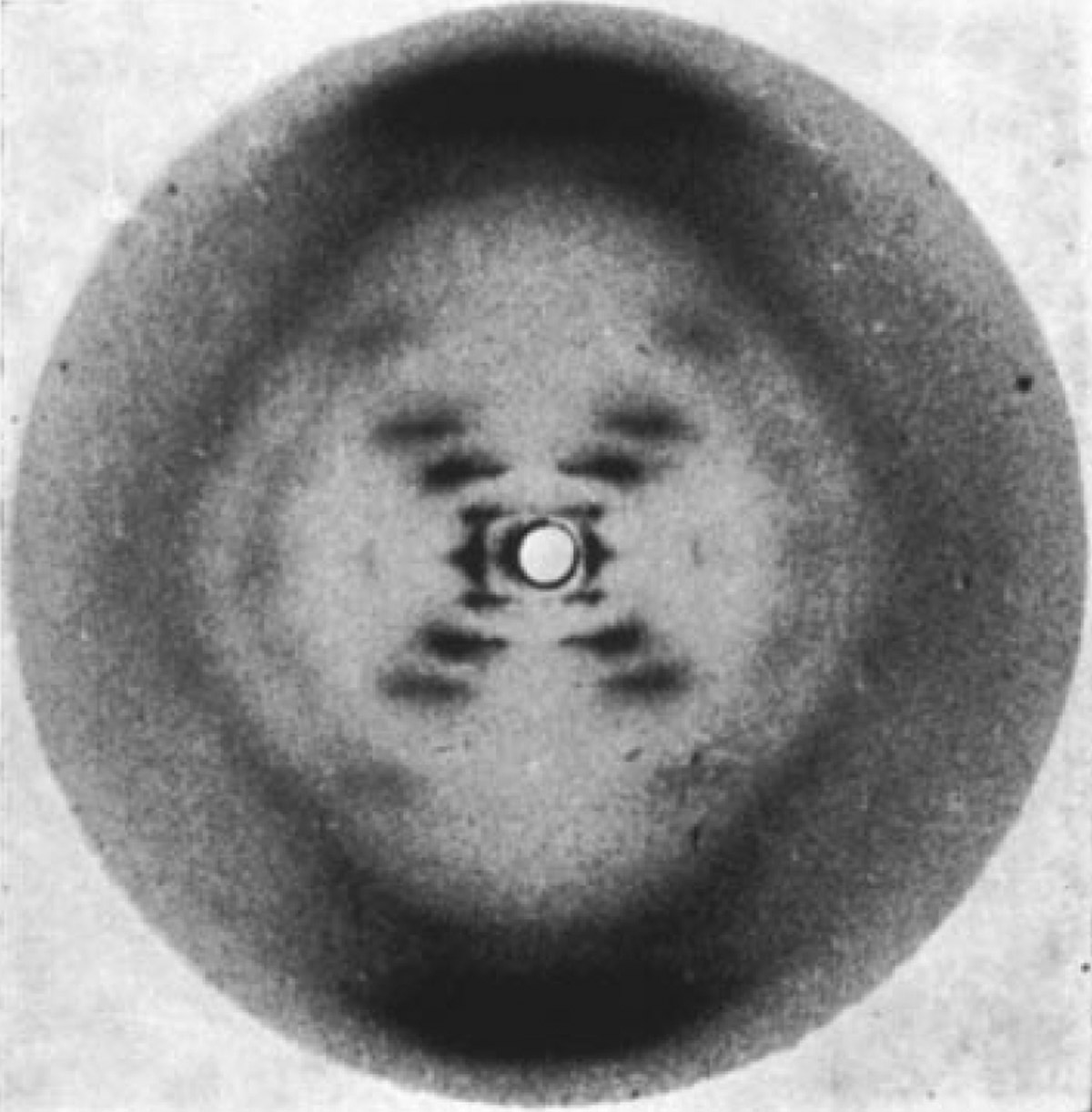
Photo 51, the B form, May 1952. From ref. 4
According to her notes she was close to solving the B structure by the end of 1952, but fed up with the mysoginistic culture in the lab, she accepted a new position studying viruses with John Bernal at Birkbeck College and was busy writing up her findings on the A form.
While Franklin and Wilkins were trying to determine the structure experimentally others turned to theoretical model building. Linus Pauling at Caltech, who predicted the α-helix of proteins with a paper cutout (winning the Nobel Prize as a result) was confident that he could solve the structure of DNA with this approach. His 3-strand model, however, would prove famously wrong.5
Meanwhile at Cambridge, the grad students James Watson and Francis Crick decided to beat Pauling at his own game but their mid-1953 theoretical model was so bad that Franklin (who had no use for model building) literally laughed at them. The problem was no one had Franklin’s photo – but that was about to change.
In early 1953, with Franklin’s departure from King’s imminent, Wilkins, without her knowledge or consent, showed Watson and Crick her image of the B form – photo 51. It was, as Watson later wrote, a revelation:
“I was shown Rosalind’s X ray photograph and ‘Whooo! That was a helix!“... I knew roughly what it meant and it was that the Franklin photograph was the key event psychologically, it mobilized us back into action.”
Using her photo and her measurements the pair went back to model-building, literally moving cardboard cutouts around on their desks. They soon determined that DNA was an antiparallel double helix with a 34 Å repeat. Within a month – on 28 February 1953 – they had figured out the structure and began work on their famous aluminum model:
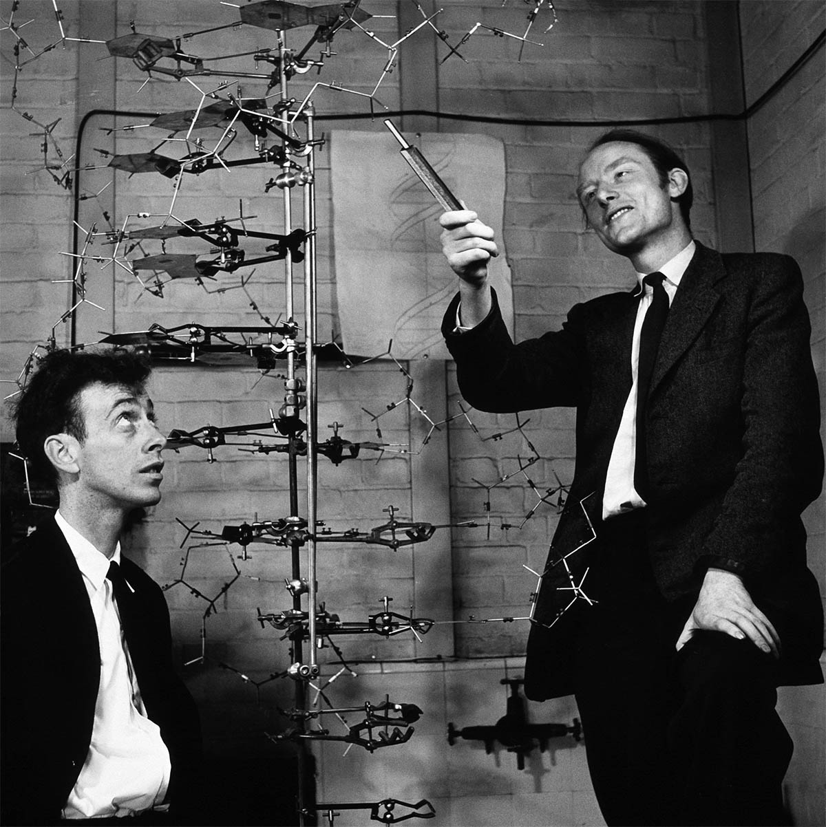
Antony Barrington Brown. James Watson and Francis Crick, 1953
In what has to be one of the most important issues of a scientific journal ever published, short papers from Watson and Crick, Wilkins, et. al. and Franklin and Gosling all appeared together in the 25 April 1953 issue of Nature.6 Of course it would be the one-page Watson and Crick paper that would be remembered.
Ironically, a lifetime of X-ray exposure and perhaps a mutated BRCA gene led to ovarian cancer for Franklin.7 She died in 1958 at the age of 37. Four years later Watson, Crick and Wilkins were awarded the Nobel Prize in Medicine for their work on DNA. That same year Pauling won his second Nobel – this time the Peace Prize.
Would Franklin have shared in the Prize were she still alive? That’s impossible to know, but according to the unsealed Nobel records Rosalind was never even considered for a nomination.
1. Avery, O.T., et. al. Studies on the Chemical Nature of the Substance Inducing Transformation of Pneumococcal Types. Journal of Experimental Medicine. 1944 Jan; 79: 137–158 (online).
2. Astbury W.T. X-Ray Studies of Nucleic Acids. Symposia of the Society of Experimental Biology. 1947; 1: 66–76 (online).
3. For a complete biography see: Maddox, Brenda. Rosalind Franklin: The Dark Lady of DNA. New York: HarperCollins, 2003 (WorldCat).
4. Attar, N. Raymond Gosling: the Man who Crystallized Genes. Genome Biology. 2013; 14: 402 (online).
5. Pauling. L., Corey, R. A Proposed Structure for the Nucleic Acids. Proceedings of the National Academy of Sciences. 1953 Feb: 39: 84-97 (online).
6. Watson J.D., Crick, F.C. A Structure for Deoxyribose Nucleic Acid. Nature. 25 Apr 1953; 171: 737-738 (online, annotated). Wilkins M.F., Stokes A.R., Wilson H.R. Molecular Structure of Deoxypentose Nucleic Acids. Nature. 25 Apr 1953; 171: 738-740 (online). Franklin R., Gosling R.G. Molecular Configuration in Sodium Thymonucleate. Nature. 25 Apr 1953; 171: 740-741 (online).
7. AKA the early-onset breast/ovarian cancer gene. This was the mutated gene Angelina Jolie discovered she had, resulting in her elective double-mastectomy. For more details see the Gene Card for BRCA1 and BRCA2, or, alternatively, any celebrity gossip website on the internet.
1 Jan 2014, updated 2 Jun 2014 ‧ Photography